Collie Eye Anomaly – CEA
30/04/2024
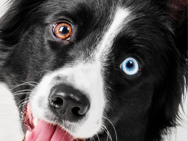
Collie Eye Anomaly (CEA) is a recessive disease that manifests through varying levels of visual impairment, ranging from no significant changes to complete blindness in affected dogs.
Generally, the two main lesions associated with CEA are choroidal hypoplasia and coloboma. Choroidal hypoplasia is characterized by a defect in the development of the choroid, which is a highly vascularized layer of the eyeball wall, located between the white part of the eye, the sclera, and the retina. Coloboma, on the other hand, is a deformation that alters the structure of the eye; in CEA it most commonly occurs in the optic nerve, where the nerve tissue is not fully developed, usually at the point where the nerve attaches to the back of the eyeball.
Cervical encephalopathy (CEA) can be diagnosed through an ophthalmological examination performed by a veterinary ophthalmologist, ideally before 12 weeks of age. After this period, clinical diagnosis becomes much more difficult due to increased iris pigmentation. Therefore, CEA can also be identified before the dog develops any clinical changes through DNA testing. The only way to prevent it is through proper breeding pairs, based on the results of genetic testing, which will prevent the birth of dogs that develop the disease.
Puppies at high risk of visual impairment (VI) can receive specialized training from an early age to learn to rely less on sight. In cases where the signs of VI are already altering the affected dog's routine, this type of training is even more important, as it will help exercise the sensory system, thus minimizing the deficit caused by vision impairment. This professional can also advise the owner on the environment and interaction with the visually impaired dog.
Roberta Costa
Veterinary Doctor
Consultant at Petgenoma



